1. The Shift to Real-Time 3D Intraoperative Imaging
Historically, surgeons relied on preoperative MRI or CT scans to build a mental map of the surgical field. However, once an incision is made, cerebrospinal fluid drains, tissues retract, and tumors are debulked, leading to a phenomenon known as "tissue shift" or "brain shift." This renders preoperative images anatomically inaccurate within minutes of opening the operative site.
Modern surgical suites overcome this through 3D intraoperative imaging (such as the O-arm or intraoperative MRI). These systems acquire updated, high-resolution volumetric datasets in real-time, feeding directly into the robotic navigation platform to ensure the surgeon is operating based on the patient's current, exact anatomical reality.
2. Core Robotic Navigation Pillars
To examine the foundational safety protocols applied in robotic-navigated surgical suites without requiring horizontal table scrolling on mobile screens, essential clinical pillars are structured into responsive cards below:
Clinical Standard: Utilizing optical or electromagnetic trackers to synchronize the patient's physical anatomy with the 3D digital model in real-time.
Clinical Standard: Creating virtual "no-fly zones" around critical structures, triggering robotic resistance if the instrument strays beyond safe resection margins.
Clinical Standard: Overlaying live fluoroscopic images onto pre-acquired 3D datasets to verify instrument trajectory without requiring continuous, high-dose radiation.
Clinical Standard: Achieving R0 resection (microscopically negative margins) by relying on millimeter-accurate spatial coordinates rather than visual estimation alone.
Journey with Confidence
Your health and well-being are our priority. Experience world-class medical excellence while exploring the wonders of Istanbul.
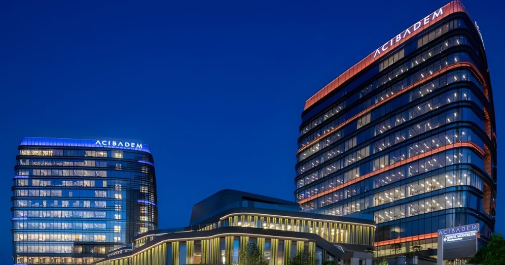
Acıbadem Hospitals Group:
Global Excellence in Healthcare
While you immerse yourself in the rich history of Istanbul, your well-being is protected by a future-focused healthcare leader. Acıbadem provides world-class medical services, from comprehensive health screenings to advanced treatments, all delivered with a commitment to your safety and comfort.
Travel with the assurance that you and your loved ones are in the most capable hands.
 Explore Medical Services →
3. Image Registration and Fluoroscopic Guidance
Before a robotic arm can safely assist in surgery, the digital space must perfectly align with the physical space. This process, known as stereotactic registration, matches anatomical landmarks on the patient (often using fiducial markers) with the corresponding points on the 3D scan. A sophisticated optical tracking camera oversees this alignment continuously.
Once registered, intermittent fluoroscopy is utilized to confirm instrument trajectories. Rather than relying on live, continuous X-ray—which exposes the patient and surgical team to heavy radiation—the system merges a single fluoroscopic snapshot onto the 3D model. This "fluoroscopic merge" guides the robotic end-effector perfectly into position for biopsy, screw placement, or tumor ablation.
4. Defining Robotic Arm Precision Thresholds
The core advantage of robotic navigation is its rigid precision. While the human hand is subject to physiological tremors and fatigue, a navigated robotic arm operates with a spatial accuracy threshold of less than 0.5 millimeters. During a procedure, the surgeon guides the instrument, but the robot enforces the mathematical boundaries.
If the surgical plan dictates that a tumor is exactly 2 millimeters away from the carotid artery, the robotic software constructs a virtual boundary. Should the surgeon’s instrument approach this boundary, the robotic arm will provide haptic resistance or physically lock, actively preventing the breach of the critical neurovascular bundle. This synergy of human decision-making and mechanical enforcement is the pinnacle of modern operative safety.

5. Maximizing Resection Margins and Neurovascular Safety
In surgical oncology, survival rates are heavily dictated by achieving an R0 resection—the complete removal of a tumor with microscopically negative margins. However, attempting radical resection near eloquent brain tissue, the spinal cord, or major vascular hubs naturally induces surgical hesitation to avoid catastrophic morbidity.
By leveraging 3D intraoperative updates and robotic navigation, this hesitation is replaced with calculated confidence. Surgeons can aggressively pursue the outermost margins of a malignancy, knowing that the navigation system will track their exact position relative to invisible, deep-seated nerves and vessels. This technology simultaneously maximizes the oncological cure rate while preserving the patient's functional quality of life post-surgery.
Tissue deformation is a major risk factor during open surgery. High-end robotic navigation systems utilize advanced elastic deformation algorithms to predict and adjust coordinates mathematically in real-time, bridging the gap between initial registration and current anatomy even before a new 3D scan is taken.
Enrich Your Exploration
Enhance your medical journey with our hand-picked partners offering luxury stays and seamless travel in Turkiye.

Four Seasons:
Bosphorus, Perfected
Escape the city's vibrant pulse to a serene waterfront palace. Here, timeless elegance and flawless service create your perfect moment.
 Book Your Escape →

Garenta:
Discover More of Turkiye
Don't just see the landmarks; experience the real culture. Garenta offers the freedom to discover hidden treasures at your own pace.
 Get Your Car →
Academic References & Data Sources
- Journal of Neurosurgery: The Impact of Real-Time 3D Intraoperative Imaging on Tumor Resection Margins and Patient Outcomes.
- International Journal of Medical Robotics and Computer Assisted Surgery: Haptic Feedback and Virtual Boundaries in Robotic Surgical Navigation.
- Annals of Surgical Oncology: Optimizing R0 Resection Rates Through Advanced Stereotactic Registration and Fluoroscopic Synergy.